

Nonlinear Optical Mineralogy (NOM)
Developing novel multiphoton microscopy applications for planetary materials


We are the first group dedicated to exploring methodological and technological development of multiphoton microscopy for non-destructive, 3D microanalysis of mineralogic samples. Multiphoton microscopy has revolutionized biomedical imaging techniques since the turn of the century and holds the potential to similarly impact fields of geologic and planetary sciences. As a unique interdisciplinary team of planetary geologists and optical scientists, we are working together to develop this powerful and versatile new toolset for targeted investigation of planetary and geologic materials.
Multiphoton microscopy utilizes a femtosecond laser to stimulate nonlinear optical interactions, wherein multiple photons simultaneously interact at the focal point of the laser to excite an electron. As the electron relaxes back to its ground state, a single photon is emitted with the summed energies of incident photons, less any energy lost to vibrational decay. The resulting signals are higher frequency (i.e., shorter wavelength) than the incident photons from the laser, and can be used to deduce structural, chemical, and electronic characteristics of minerals.
Nonlinear optical interactions are extremely unlikely to occur outside of the laser's focal volume, only occurring at detectable levels where the flux of photons is exceptionally high. We can take advantage of this by using incident laser wavelengths for which the sample is largely transparent to collect images and emission spectra at-depth. Such images can then be stacked in a depth profile to reconstruct 3D nonlinear optical renderings, showing minimally aberrated structural and fluorescent details of crystalline interiors.
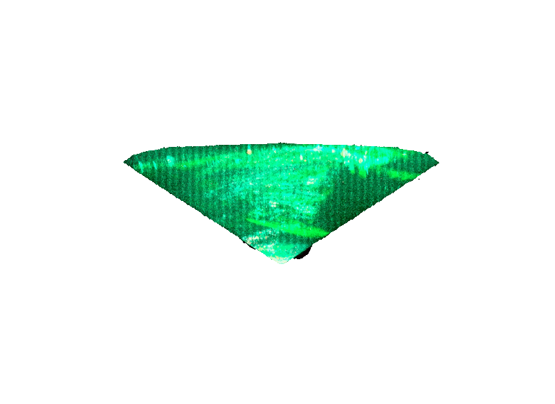
An example of a faceted pink diamond (2 mm diameter) from the Argyle mine in Western Australia is shown below in false color. In this image, green diagonal bands are produced by 3-photon (i.e., third harmonic, THG) generations where a change in refractive index occurs. These are dislocation slips, formed during high-pressure deformation events as the diamonds ascended through the crust in a kimberlite dike. Blue/cyan fluorescence is also present throughout its volume; spectral analysis shows that fluorescence arises from N3 vacancy centers. These centers accumulate in diamonds over long periods of time at high temperature, supporting the natural provenance of this gemstone. Other mineral inclusions can also be seen in irregular green THG shapes, as well.
Current NOM Group Members


Related Research by Topic
Nonlinear optical mineralogy and the search for water in the early solar system:
Multiphoton microscopy applications for astromaterials (conference abstract)
Multiphoton microscopy as a novel tool for investigating evidence of aqueous activity in chondrites (conference abstract)
Harmonic identification of extrasolar minerals:
Rapid detection of silicon carbide using multiphoton microscopy (conference abstract)